
Very few things strike as much fear in most EMS providers more than the critically ill or
injured pediatric transport. Some of the forces that instill fear in most EMS providers is the
lack of available appropriately sized pediatric equipment, the general lack of familiarity with
what would be deemed “normal” vital sign parameters, the anatomic and physiologic
differences between the adult and pediatric population and the general approach to the
assessment of the pediatric patient.
Physiologically, pediatric patients are faster metabolizers of oxygen (often at a rate of
2-3 times that of an adult), they have smaller oxygen reserves and small airways. This makes
them more prone to desaturation and complete or partial airway obstructions. Their accessory
ventilation muscles are less developed and ribs are more cartilaginous and horizontal in nature
which makes their negative pressure spontaneous ventilation less effective and the diaphragm
is more important for adequate ventilation. Overall the young pediatric patient’s airway is funnel
shaped with the narrowest portion slightly below the glottic opening while their adult
counterparts have a more cylindrical shaped airway with the narrowest portion at the glottic
opening. Couple that with a large, floppy epiglottis and large tongue in relation to their overall
oropharynx can make managing the pediatric airway much more intimidating. Positioning of
the airway is extremely important when managing the pediatric airway. Often hyperextension
of the head can occlude the very narrow and flexible trachea of young pediatric patient.
Padding becomes very important to maintain proper airway alignment. Pediatrics have less
developed compensatory mechanisms that can lead to rapid deterioration in the presence of
hypo perfusion. They rely on increasing their stroke volume more than their ability to increase
stroke volume. They tend to be able to maintain a “normal” blood pressure longer by shunting
blood to the core due to vasoconstriction of peripheral blood vessels but when they
decompensate, they decompensate rapidly. Renally speaking, their immature kidneys are less
effective in concentrating or diluting urine when necessary so they are more prone to volume
depletion and/or volume overload and electrolyte abnormalities. Cartilaginous bone structures
of the pediatric patient are less likely to absorb traumatic energy and more likely to allow that
energy to pass through the bone structure without fracturing and injuring the organs and blood
vessels below.
Vital sign parameters vary widely among pediatric patients and heart rate and
respiratory rate trend down as the patient grows older while the overall blood pressure and
Mean Arterial Pressure (MAP) tend to increase with age. A helpful formula to estimate an
acceptable systolic blood pressure in the child between 1-10 years of age is (Age in Years x 2)
+ 70. Anything below this number is considered hypotensive. A systolic blood pressure of
approximately 70 mm/Hg is considered normal in infants less than 1 year of age and a systolic
> 90 mm/Hg is considered normal in patients older than 10 years old. Normal urine output for
the pediatric patient less than 8 years old or under 30 kg’s is 1-2 ml/kg/hr while pediatric
patients older than 8 years of age and greater than 30 kg have a normal urine output of 0.5-1
ml/kg.
Assessment of the pediatric patient can be difficult and often the Pediatric Assessment
Triangle is utilized to determine the severity of their medical or traumatic injury or condition.
The PAT is an “across the room” assessment that is done rapidly and without actually touching
the patient. The 3 sides to the Pediatric Assessment Triangle are A=Appearance, B=Work of
Breathing and C=Circulatory Status. The appearance evaluation is based on the TICLS
mnemonic (T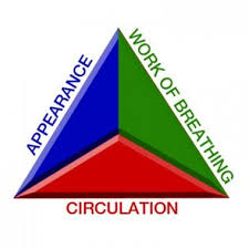 one, Interactiveness, Consolability, Look or Gaze and Speech or Cry) Appearance
involves overall positioning of patient and if they are reacting to their environment in an age
appropriate fashion. Work of Breathing involves the presence or absence of adventitious lungs
sounds or audible respiratory sounds, accessory muscle use and overall adequacy of
ventilatory rate and volume. Circulatory assessment involves determination of skin color,
condition and perfusion status. If all 3 sides are normal, the patient is often considered “Not
Sick”, if 1 side is abnormal than the patient is considered “Sick”, if 2 sides are abnormal than
the patient is considered “Critical” and if all 3 sides of the triangle are abnormal than the
patient is considered to be in cardiopulmonary failure.
Because true pediatric emergencies are rare in most EMS providers’ daily routine, they
tend to evoke much higher anxiety levels in most providers. Understanding the anatomic,
physiologic, vital sign parameters and assessment technique differences when dealing with the
pediatric patient will help the provider to feel more comfortable treating the younger patients. It
can also allow us to feel more comfortable when interacting with them and providing
competent, complete and consistent care to our youngest patients.