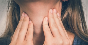
A variety of triggers can cause a laryngospasm, such as asthma, allergens, liquid, dust, fumes, infection, smoke, foreign body, etc. Other causes included hypocalcemia, vagal hypertonicity or painful stimuli.
Authorities define laryngospasm as as an uncontrolled or involuntary muscular contraction of the vocal cords and ligaments. The vagus nerve has actually proven a predominant cause of nervous mediation. The superior laryngeal and pharyngeal branch of C Nerve X (CN X) and the recurrent laryngeal compose the vagus nerve. The condition effects the cricoarytenoid, thyroarytenoid and the lateral cricoarytenoid.
According to Open Anesthesia, the larynx itself is composed of nine cartilages — three paired and three unpaired — that contain the vocal cords within them. The extrinsic muscles that move the larynx as a whole and the intrinsic muscles that move the various cartilages in relation to one another control the movements of the larynx.
It becomes innervated bilaterally by the superior laryngeal nerve, which supplies mucosa from the epiglottis to the level of the cords, and the recurrent laryngeal nerve, which supplies mucosa below the cords. Both are branches of the vagus nerve.
The recurrent laryngeal nerves supply all the intrinsic muscles of the larynx except for the cricothyroid muscle. Any damage to the recurrent laryngeal can result in vocal cord dysfunction, according to Open Anesthesia. The external branch of the superior laryngeal nerve innervates the cricothyroid muscle.
Other nerves include the maxillary branch of the trigeminal nerve that supplies sensory innervations to the nasopharynx and the glossopharyngeal nerve that supplies sensory innervations to the post one-third of the tongue, pharynx and areas above the epiglottis.
Protecting the airway from aspiration
When reflexes are an issue
Both the gag reflex and the aerodigestive play an important role in mediating laryngospasm and avoiding aspiration by responding to irritants — objects and liquids in the vicinity of the epiglottis, glottis, larynx and the upper esophageal sphincter — with expulsion from the area of the irritant, object or liquid.
Distinct yet inter-related, these two reflex mechanism serve to protect the airway from aspiration. In essence, swallowing triggers reflexes in the larynx, epiglottis and esophagus that effectively isolates the airway.
Because of the anatomic contiguity between the pharyngolaryngeal and gastroesophageal pathways, these reflexes function during retrograde (reflux) passage of stomach contents as well as antegrade (swallowing) passage of substance via the digestive tract protecting the airway from aspiration.
Several aerodigestive reflexes triggered at various levels have been proposed to protect the airways against aspiration, according to an article in Gastroenterology. Distention of the esophagus, for example, can enhance upper esophageal sphincter pressure — called the esophago upper esophageal sphincter contractile reflex — and this may prevent entry of esophageal contents into the pharynx.
When fluids are an issue
 Fluid in the pharynx can enhance upper esophageal sphincter pressure — called the pharyngo-upper esophageal sphincter contractile reflex — and may protect against further esophagopharyngeal reflux.
At a larger volume, fluid in the pharynx also can trigger an irrepressible swallow called the reflexive pharyngeal swallow that not only triggers glottal closure, but also clears the pharynx of any fluid, according to articles in Gastroenterology, Anesthesiology and Gut.
Glottal closure without a swallow also can be triggered by fluid in the pharynx called the pharyngoglottal closure reflex, according to articles in Gastroenterology, Gut and Gerontology.
The Gastroenterology article reports that though researchers have proposed that these aero-digestive reflexes protect the airways against aspiration, they’ve found no evidence to directly show their role in airway protection.
Effecting glottic closure via the aerodigestive reflexes precludes laryngospasm. However there’s a limit to the volume of fluid that can remain in the hypopharynx before spilling into the larynx.
This suggests that receptors triggering reflexive pharyngeal swallowing reflex may be located at the area near the upper margin of the interarytenoid fold and are not amenable to volitional suppression of swallow as are those receptors located in the rest of the pharyngeal swallow trigger zone such as the posterior pharyngeal wall or tonsillar pillars. This results in triggering reflexive pharyngeal swallowing when accumulated fluid reaches that area in otherwise healthy individuals with intact sensations.
Laryngospasm causes
There are many causes of laryngospasm, however many often occur during anesthesia. Specifically during induction and recovery, such as under light sedation. This includes laryngopharyngeal reflux during induction or if the patient is under sedated.
Additionally, inadvertent extubation tends to precipitate laryngospasm. Judging from the bulk of the literature, the problem lasts from seconds to a few minutes and eventually resolves spontaneously.
Reports on this information come primarily from surgical settings, most often elective surgery. In such instances de-saturation become less of a problem than in the pre-hospital setting where hypoxia may arise.
Spontaneous resolution of laryngospasm refers to cases of sleep apnea or episodes of gastric reflux, which often occur during sleep. CPAP has become a popular treatment for the former. Treatment for the latter now focuses on relieving the underlying cause of the gastric esophageal reflux disease (GERD). Episodes of laryngospasm can last 20 to 30 minutes necessitating aggressive treatment.
Research on laryngospasm treatment options
Treatment focuses on relaxing the laryngeal muscles and tendons.
In instances of laryngopharyngeal reflux and surgery, treatment would continue expeditiously to paralysis, similarly in surgery, to re-anesthetize the patient. Thus, it appears there is some higher cortical involvement in alleviating the reflex.
Research on puppies vs. adult dogs demonstrated that laryngospasm was more prevalent in the puppies before maturity, reported in the article “Effect of bilateral vagosympathetic nerve blockade on response of the dog upper esophageal sphincter (UES) to intraesophageal distention and acid” that appeared in the journal Gastroenterology. The implication of this is borne out in clinical practice because the condition is more common in pediatric patients.
Researchers think higher cortical involvement plays at least a partial role in the spontaneous resolution of such episodes. Experts often associate the cause under light anesthesia with some stimulus to the areas innervating the gag and aerodigestive reflexes.
Lighter anesthesia may impair these protective mechanisms where as full sedation or paralysis completely inhibits them. Chemical intervention is the treatment of choice in these circumstances if it is readily available.
“Aggressive jaw thrust and forceful ventilation” has become the generally accepted immediate approach to resolving laryngospasm. That means a good airway maneuver and effective positive pressure ventilation.
Laryngospasm treatment in the field
Medical professionals use this approach with drowning victims where patients present persistent laryngospasm. The logic is to attempt to force air against and ultimately through the larynx. This may be moot if concomitate epiglottic spasms occur as well.
Ideally, a proper jaw thrust will at least move the epiglottis out of the way allowing PPV against the larynx. Partial hypoxia can account for the spasm via impairment of higher cortical involvement as well as reflex mechanisms mentioned. Complete and profound hypoxia will cause relaxation of the muscles, but this is not a recommended treatment method.
One method was put forth by Phil Larson called the Larson Maneuver, according to the article “Laryngospasm — The Best Treatment” published in the journal of Anesthesiology. Some literature describes it as anticandidal, however, according to Larson it works every time.
The maneuver involves pressure on the “laryngospasm notch” or “Larsons Point” in conjunction with a jaw-lift maneuver. This technique may involve vagal or painful stimulation leading to easing of the spasm.
“This notch is behind the lobule of the pinna of each ear,” Larson said. “It is bounded anteriorly by the ascending ramus of the mandible adjacent to the condyle, posteriorly by the mastoid process of the temporal bone, and cephalad by the base of the skull.”
