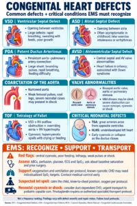
Congenital heart defects (CHDs) are the most common type of birth defect and affect approximately 1% of newborns in United States. A congenital heart defect is defined as a structural or functional problem of the heart or great vessels that is diagnosed prior to or present at birth. CHDs vary in severity from barely perceptible at birth to life threatening to the point that pre birth surgery is required to ensure fetal viability. There are many forms of congenital heart defects but the most common way to classify them is based on how the defect affects blood flow and functionality of the heart. Today we are going to look at cyanotic and non cyanotic (acyanotic) CHDs.
Acyanotic or non cyanotic heart defects result in a left to right blood flow shunt pattern within the heart itself. Remember that the right side of the heart contains less oxygenated blood while the left side of the heart pumps the oxygenated blood to the systemic circulation. Because non cyanotic CHDs result in this left to right shunt pattern, the symptoms often appear less apparent and non cyanotic CHDs frequently are less dangerous to the neonate.
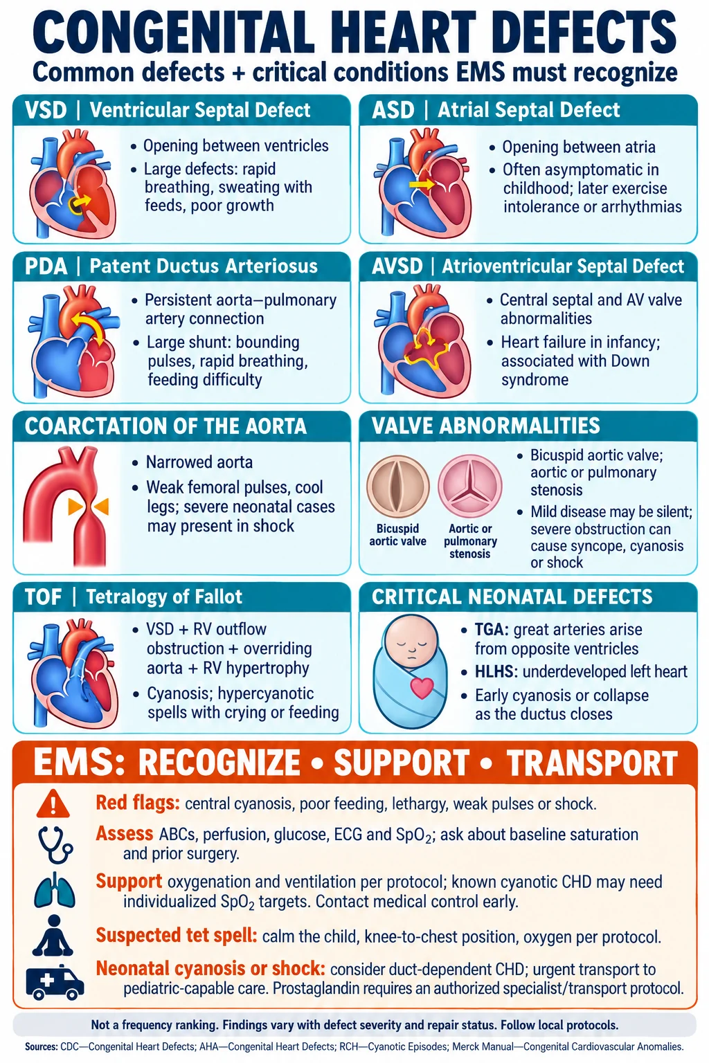
These neonates often present with mild to moderate congestive heart failure, poor exercise and feeding tolerance, poor weight gain and extended recovery from simple respiratory infections. The most common non cyanotic CHD is a ventricular septal defect or VSD and accounts for around 20-25% of all defects. It is basically a hole in the septum of the ventricles that allows the more oxygenated blood to pass from the left to the right ventricle due to the higher pressure gradient of the left ventricle. Atrial septal defects or ASDs are the second most common CHD accounting for approximately 10-15% of all CHDs. This results in hole that blood is pumped through from the left atrium to the right atrium. Minor ASDs are often only recognized during post delivery exam when a heart murmur is discovered while more severe ASDs may require surgical intervention early in life to correct the malformation. Pulmonary stenosis is a narrowing of the pulmonary valve or the blood vessel beyond it that limits blood flow exiting from the right ventricle to the lungs. Pulmonary stenosis accounts for approximately 10% of all CHDs and presentation will vary based on the severity of the narrowing.
Cyanotic CHDs will result in a right to left shunt of blood flow in the heart and often presents with more significant signs and symptoms due to the fact that the less oxygenated blood is being shunted from the right side of the heart to the left side and then it is pumped throughout the body. The patient will often present with more severe exercise intolerance (when feeding or crying), cyanotic spells, apneic spells especially when the neonate is under stress, inconsolable irritability, diaphoresis and in severe cases may result in in seizures, coma or cardiac arrest. Tricuspid atresia is a structural disorder in which the heart has only 3 functional valves rather than 4 as the tricuspid valve doesn’t open. This defect will not allow the right atrium to eject its blood to the right ventricle. Emergency surgery is often indicated for tricuspid atresia unless the patient has an atrial septal defect that will serve as an auxiliary pathway for blood to exit the right atrium. Hypoplastic left heart syndrome results in a smaller, functionally inadequate left ventricle that often necessitates surgery within the first few days of life and repeated follow up surgeries as the child develops with age. Transposition of the great arteries accounts for approximately 5% of all CHDs. The aorta is connected to the right ventricle and pulmonary artery is connected to the left ventricle therefore delivering less oxygenated blood to the body and more oxygenated blood to the lungs for an additional pass. Tetrology of Fallot is actually not just one congenital heart defect but actually 4 defects that occur to some degree simultaneously. The neonate has a large ventricular septal defect combined with right ventricular stenosis or narrowing, an enlarged aorta that sits directly above the large VSD and an overall enlargement or thickening of the wall of right ventricle. Survival rates for Tetrology of Fallot is dictated by the degree of the CHD as well as the presence of serial surgical interventions that must occur during the lifespan of the patient due to growth patterns and supply/demand imbalances. These patients often require life long monitoring and often suffer from other congenital defects both within the cardiovascular system as well as other organ systems.
While no clear consistent root is cause is known for congenital heart defects, there are some contributing factors that increase the likelihood of a CHD developing during gestation. Genetic factors, pre-existing maternal health disorders, substance use during pregnancy and other environmental exposures have been thought to contribute to their development. Early recognition coupled with well performed treatment modalities have been shown to improve both short and long term outcomes. Many patients have the potential to lead healthy, long lives even with severe CHDs if these disorders are aggressively treated and disorder progression is monitored.