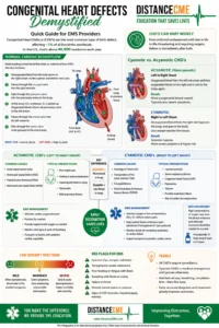

Congenital Heart Defects (CHD’s) are the most common type of birth defect and can
affect approximately 1% of all live births worldwide. In the United States, this adds up to about
40,000 newborns annually. Congenital heart defects have varying degrees of severity and
many can be overlooked and/or diagnosed later in life. The term “congenital” means the
condition was present at or before birth, whereas “acquired” usually describes a condition that
has been obtained or developed over time. With today’s technological advances, many CHD’s
can be diagnosed and sometimes repaired in utero, before the baby is born. CHD’s can be
roughly divided into Cyanotic and Acyanotic (or noncyanotic) in nature. Acyanotic CHD’s result
in a left to right shift in blood flow through the chambers of the heart while cyanotic CHD’s
cause a right to left shift in blood flow patterns.
In order to understand the differences and significance of these two heart disorders, it’s
important that we review normal blood flow as it passes from the inferior and superior vena
cava to the aorta. Normal cardiac blood flow enters the right atrium from the inferior and
superior vena cava as it returns from the systemic circulatory system. The right atrium ejects
the less oxygenated blood through the tricuspid valve into the right ventricle which then ejects
blood through the pulmonic valve into the pulmonary artery which then divides to allow the
blood to travel to each lung. Once the blood arrives at the lungs, CO2 is diffused across the
alveolar capillary membrane for exhalation and inhaled O2 is picked up by the hemoglobin in
the pulmonary blood vessels to be returned to the left atrium via the pulmonary veins. The
oxygenated blood leaves the left atrium via the mitral valve and is then pumped throughout the
entire body as it leaves the left ventricle via the aortic valve.
With Acyanotic CHD’s, there is a hole, usually somewhere in the septum of the heart,
that allows the already oxygenated blood from the left side to mix with the less oxygenated
blood of the right side. This mix of oxygenated and less oxygenated blood is then pumped to
the lungs again to become more oxygen rich. Because of this blood flow pattern, non cyanotic
CHD’s have a tendency to present with less severe symptomologies and usually have a better
prognosis. Acyanotic CHD babies will often present with poor weight gain, feeding or exercise
intolerance, sweating while eating or crying and sometimes respiratory distress and
tachycardia. Treatment for non cyanotic CHD’s frequently involves minimizing cardiac oxygen
demands, providing supplemental oxygen when necessary and transporting to the appropriate
facility that is equipped to evaluate and/or surgically repair the defect if necessary.
Cyanotic CHD’s on the other hand, cause a right to left shift in blood flow patterns.
Cyanotic CHD’s are usually due to a hole in the septal walls of the heart or an abnormality with
the great vessels that exit the] ventricles of the heart. This results in the less oxygenated blood
from the right side of the heart to not make it to the lungs for CO2 offloading and O2 uploading.
The less oxygenated blood then travels to the remainder of the body via the systemic
circulation and never has the chance to become oxygen rich. The blood flow deviation leads
to varying levels of systemic tissue hypoxia and can present with more severe symptoms.
Cyanotic CHD’s also have a more guarded prognosis and a tendency to have a higher mortality
and morbidity and their non cyanotic (acyanotic) counterparts. Presenting signs and
symptoms of cyanotic CHD’s tend to be more severe and can occur earlier than non cyanotic
CHD’s. Cyanosis, congestive heart failure (CHF) due to pulmonary congestion, severe
respiratory distress and sometimes respiratory failure, seizures due to hypoxemia and death
can occur. Treatment of cyanotic CHD’s is a little different. In severe cases, sub atmospheric
fractions of inspired oxygen (FiO2) around 15-20% may be required as higher concentrations
of oxygen may cause other associated defects, such as the Ductus Arteriosis, to close. The
patent ductus arteriosis often provides accessory blood flow that is necessary for survival until
repair can be performed. Administration of Prostaglandin E1 will also aid in keeping the ductus
arteriosis open until surgical repair can be performed.
As you can see, CHD’s have many varying presentations and levels of severity. The
severity ranges from barely perceptible and sometimes overlooked until later in life to life
threatening to the point that surgical repair is necessary either before the baby is born or
immediately post delivery. While all CHD’s require some level of surveillance, the more severe
forms, often cyanotic CHD’s, tend to present with more acute decompensation and the
symptoms can be more severe. Early, accurate diagnosis and treatment has been proven to
equate to a markedly lower morbidity and mortality and generally a better long term prognosis.